HUU (Hyperuricosuria and Hyperuricemia)
Written by Aurélie Allard, Animal Health Technician
(Translation DR)
HUU is a genetic metabolic disease that can occur in mammals, including humans and dogs. Hyperuricosuria indicates high levels of uric acid in the urine, while hyperuricemia indicates high levels of uric acid in the blood. Uric acid is a metabolic by-product of DNA metabolism. Notably, uric acid is not very soluble in body fluids and has a tendency to come out of solution (to precipitate), forming crystals in the blood and stones in the urinary system. It is these crystals and stones formed by precipitated uric acid that are the cause of HUU.
To prevent these problems, in most mammals, uric acid is quickly converted to another metabolic product, allantoin. Allantoin is much more soluble in body fluids than uric acid is, making it much easier for the body to eliminate in the urine. Unfortunately, in humans and great apes (and the dalmatian dog), there are problems in converting uric acid to allantoin. Uric acid levels can build up in body fluids, resulting in the hyperuricosuria and hyperuricemia that is seen in HUU.
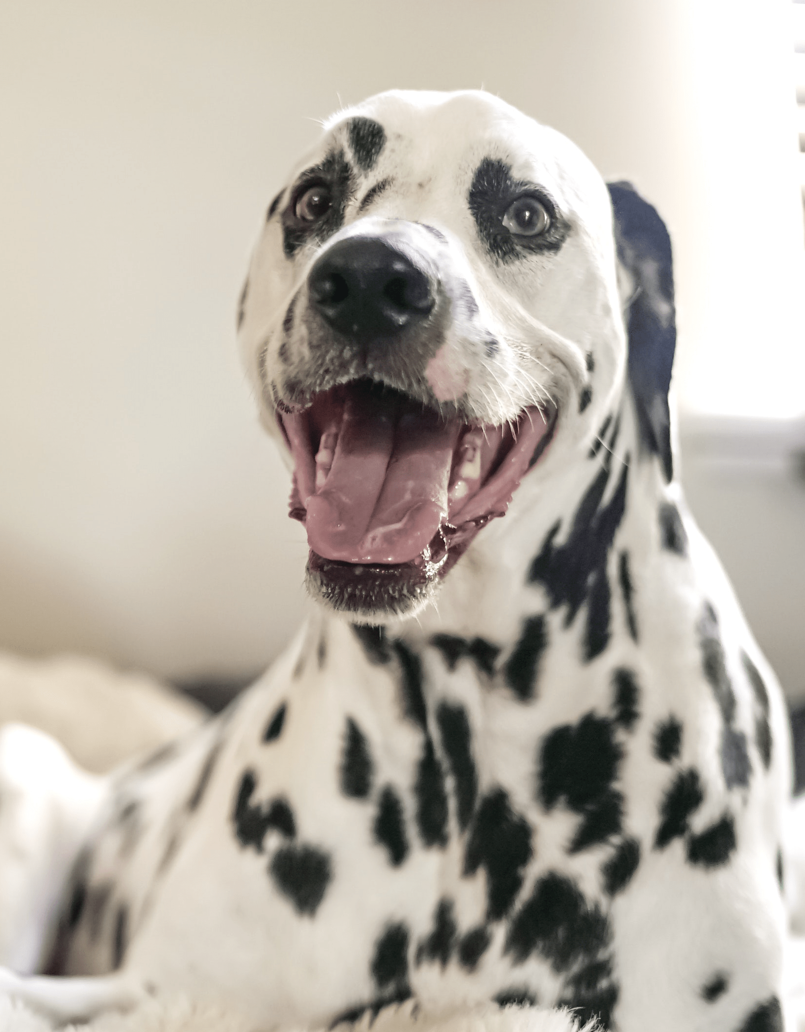
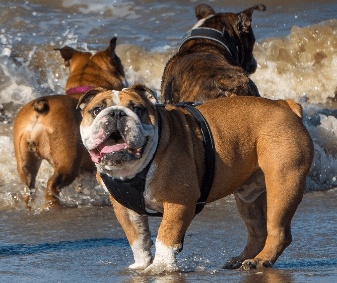
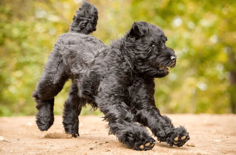
Dogs from many breeds can suffer from HUU. However, several breeds, including Dalmatians, English Bulldogs and Black Russian Terriers are particularly affected by this disease.
Clinical signs of Hyperuricosuria and Hyperuricemia
In humans, HUU can cause gout, a chronic disease that affects the joints, and also kidney and bladder stones resulting in urinary blockages. In dogs, HUU results in bladder stones and clinical signs including difficulty in urinating, frequent small volume of urination (micturition), blood in the urine, abdominal pain, lack of appetite, lethargy and vomiting. Because of their anatomy, HUU is more common and more severe in middle-aged male dogs. When a urinary blockage occurs, there is a risk of bladder rupture and even death. This is a medical emergency and veterinary intervention is required.
The Diagnosis of HUU
In dogs, the diagnosis of HUU is made based on clinical signs, with confirmation by X-rays and laboratory tests. For a dog with a urinary problem, the veterinarian will take a urine sample to check for uric acid crystals as well as blood cells. Uric acid is barely soluble in urine at body temperature. When the urine sample is cooled to room temperature, uric acid forms small crystals that are visible to the naked eye, which is an aid in the diagnosis of HUU.
The veterinarian can also take X-ray and ultrasound images of the bladder to see if there are bladder or kidney stones. There are several possible causes of bladder stones; for stones caused by uric acid, ultrasound is the most useful technique.
Treatment and Prevention of HUU
Fortunately, the symptoms of HUU can be treated and the occurrence of symptoms can be prevented. However, dogs that are susceptible to HUU will need to be monitored regularly by a veterinarian. A special diet low in purine and protein is recommended. Dehydration is to be avoided, and moist food should be considered. Surgical removal of kidney stones is an option and at times, a necessity. In all cases, in order to avoid the recurrence of symptoms, the dog should be kept on the special diet and kept well hydrated.
The Genetic Profile of Hyperuricosuria
In dogs, hyperuricosuria is an autosomal recessive genetic disease with variable penetration. The disease can affect both males and females, but is seen more frequently in males because of their long urinary tract. This genetic disease is caused by a mutation in the SLC2A9 gene, which codes for the protein that carries uric acid in the liver and kidneys. The mutation itself is a simple typo in the DNA; the substitution of a G for a T in the SLC2A9 gene.
Remember that our genetic system is duplicated; each dog has two copies of the SLC2A9 gene, one from the sire and the other from the dam. If one copy of the gene is mutated, the animal is a carrier (M/N) for the mutation but is not at risk for the disease. However, he will be able to transmit the mutated gene to his descendants. On the other hand, the double mutated animal (M/M), with both copies of the mutated gene, is at risk of having hyperuricosuria as well as passing the mutated gene on to puppies. Finally, the disease is said to be of “variable penetration”. An animal (M/M) may have a few crystals in the urine but not the disease, or may have a urinary blockage with serious consequences.
Mutation Frequencies
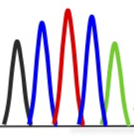
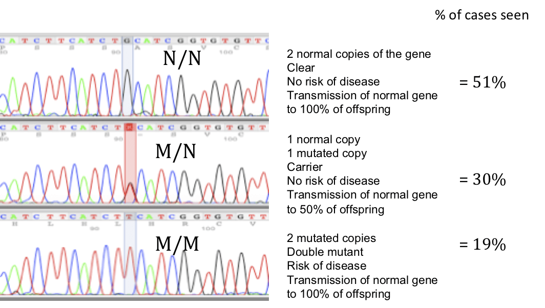
At the Laboratory of Veterinary Genetics (Labgenvet), we have tested over 200 dogs for the mutation in the gene that causes HUU. Of these dogs, 51% were clear (N/N) with 2 copies of the normal gene. 30% of animals tested were carriers (M/N) with one copy of the normal gene and one mutated copy. Finally, 19% of the dogs tested (1 in 5) were double mutated (M/M) and therefore at risk of developing hyperuricosuria during their lifetime. The DNA profiles for the N/N (clear), M/N (carrier) and M/M (double mutant, at risk) samples received and the overall percentages of these cases are shown in the illustration below:
Site of the mutation in the SLC2A9 gene causing HUU in Dogs
The HUU Mutation is an Old Mutation
The mutation responsible for HUU is found in a number of breeds of dogs. This fact suggests that the mutation is an old mutation that was present before the formation of the modern dog breeds, which occurred from 100 to 150 years ago. This was confirmed by the findings of a DNA study reported in 2017. The skeleton of a terrier-type dog was found aboard the wreck of the tall ship Mary Rose which sank in 1545. DNA analysis on the skeleton showed that the dog was a carrier (M/N) for the mutation that causes HUU.
Hyperuricosuria in Crossbreed Dogs
Hyperuricosuria is present in pedigree dogs, but it can also be found in crossbred dogs. In a study that was conducted on almost 35,000 crossbreed dogs, 4.5% were carriers (M/N) and 0.2% were double mutated (M/M) for the mutation responsible for hyperuricosuria.
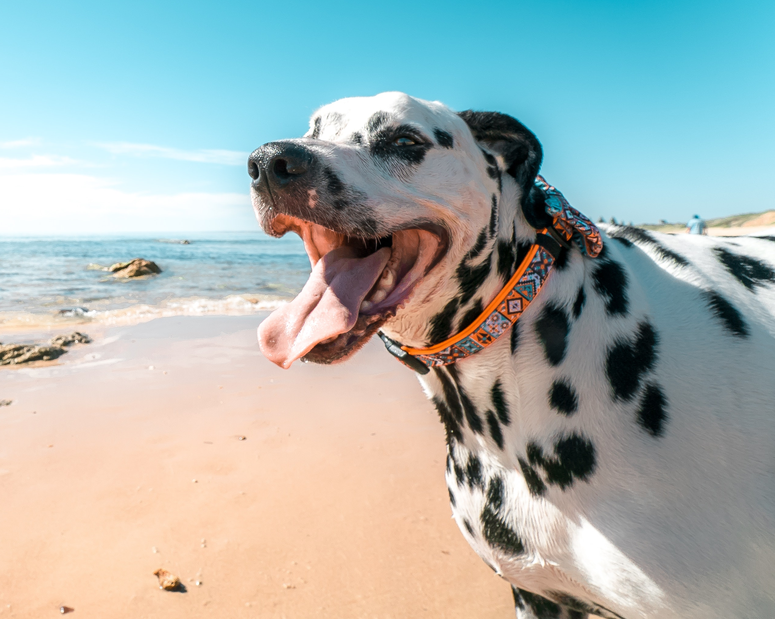
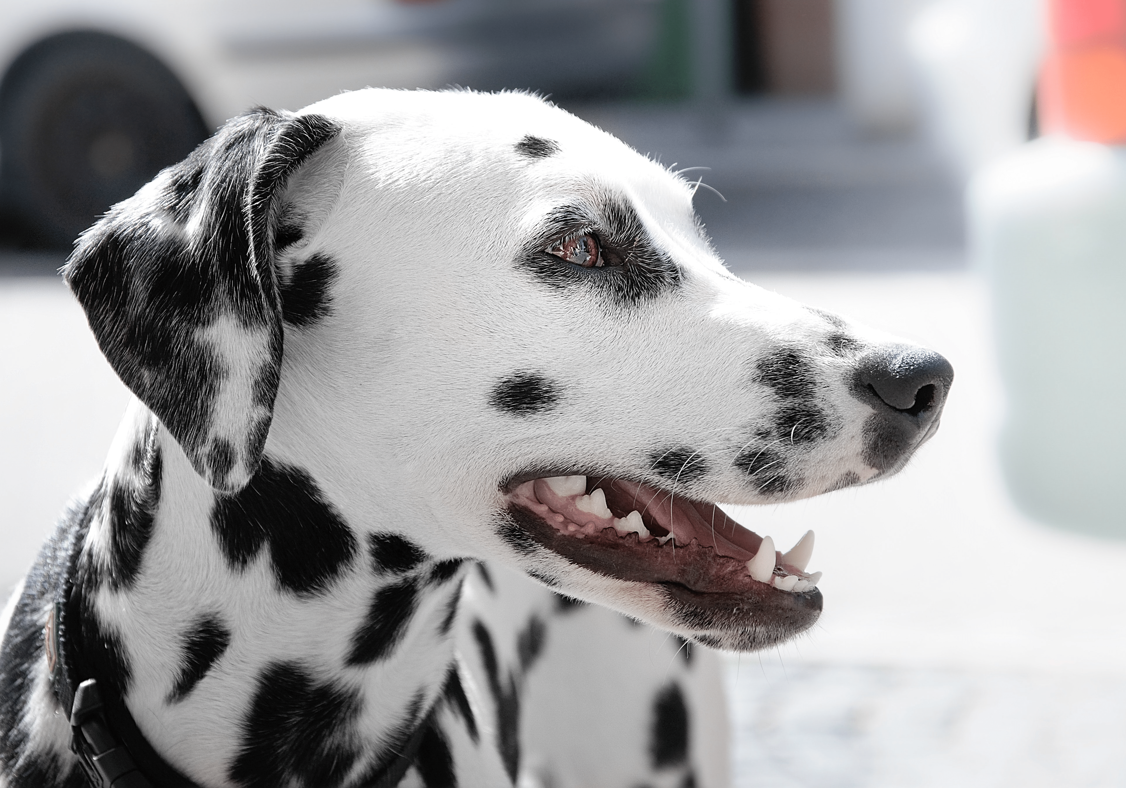
The Particular Case of the Dalmatian
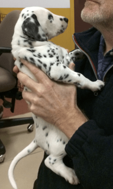
Dalmatians as a breed of dog are at greater risk of uric acid stones and developing HUU than other dog breeds. This is due to the fact that the mutation in the SLC2A9 gene responsible for HUU was inadvertently fixed in the genome of Dalmatians when the breed was formed, possibly at the same time as selecting for the distinctive black spots of their coat. In 1973, Dr. Robert Schaible crossed an English Pointer (N/N, clear for the mutation for HUU) with a Dalmatian (M/M, double mutant) in order to eventually create Dalmatians free from the genetic mutation. Now, after 50 years, Dalmatian breeders are mating descendants of this cross to initially select Dalmatians that are carriers (M/N) but not at risk for HUU, and ultimately select Dalmatians that are N/N clear for the mutation responsible for HUU.
Breeding Strategies
In order to prevent HUU in a breed or pedigree with a known predisposition, DNA testing should be done to determine the genetic status of breeding animals. The best breeding strategy is to mate two animals that are clear (N/N) for the mutation in question. Since HUU is a recessive genetic disease, an animal must have a double mutation to be at risk of the disease. Alternatively, a carrier dog (M/N), which is not at risk of the disease, but which can transmit the mutation to the next generation, can be mated with an animal that is clear (N/N). In this case, half of the litter (on average) will be a carrier for the HUU mutation, but no puppy will be at risk for the disease.
The mating to avoid is between two carrier animals (M/N by M/N), as one in four puppies (on average) will be at risk of being double mutated (M/M) and at risk of HUU. Animals that are double mutated (M/M) are not good candidates for breeding. If a double mutated dog (M/M) is identified by a DNA test at a young age, the veterinarian can immediately prescribe a special diet for his condition.
Références:
- OMIA link: [1033-9615]
- Zierath S, Hughes AM, Fretwell N et al. (2017) Frequency of five disease-causing genetic mutations in a large mixed-breed dog population (2011-2012). PLoS One 12(11):e0188543. [pubmed/29166669]
- Westropp JL, Larsen JA, Johnson EG et al. (2017) Evaluation of Dogs With Genetic Hyperuricosuria and Urate Urolithiasis Consuming a Purine Restricted Diet: A Pilot Study. BMC Vet Res. 13(1):45. [pubmed/28178975]
- Karmi N, Safra N, Young A et al. (2012) Validation of a urine test and characterization of the putative genetic mutation for hyperuricosuria in Bulldogs and Black Russian Terriers. Am J Vet Res. 71(8):909-14. [pubmed/20673090]
- Karmi N, Brown EA, Hughes B (2010) Estimated frequency of the canine hyperuricosuria mutation in different dog breeds. J Vet Intern Med 24:1337-1342. [pubmed/21054540]
- Bannasch D, Safra N, Young A et al. (2008) Mutations in the SLC2A9 gene cause hyperuricosuria and hyperuricemia in the dog. PLoS Genetics 4(11):e1000246. [pubmed/18989453]